
As the field of breast imaging has evolved over time, the role of the breast imager has as well. Historically, radiologists were perceived to be in their reading rooms all day, interpreting the imaging, issuing reports to referring providers and consulting with other physicians behind the scenes in the care of patients. Today, breast imaging radiologists play an active and critical role in the multidisciplinary team and comprehensive care of each breast cancer patient.
As we observe Breast Cancer Awareness Month and 100 years of the ACR, we reflect on where we started and where we are going in the pursuit to provide not only high-quality care but also equitable access to care for all our patients, particularly when it comes to mammography quality, access and reimbursement.
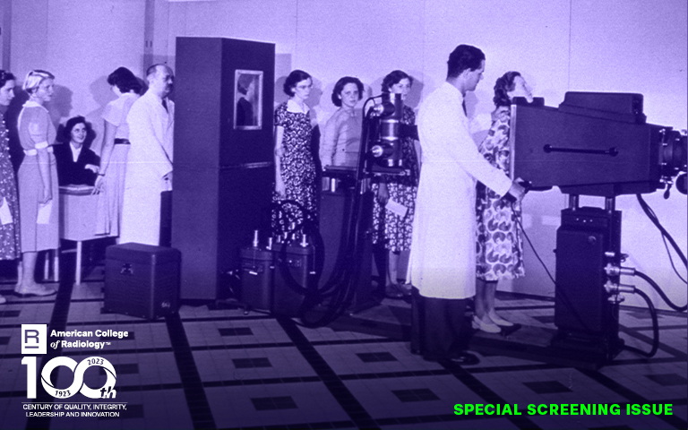
Early Breast Imaging
Mammography screening has certainly come a long way since its inception. In the 1960s, mammography was performed using a general-purpose X-ray tube with images captured on direct exposure film and developed in a darkroom. There was no compression, and the images were low in contrast resolution.
In the following decade, significant advancements were made with the introduction of screen-film mammography. This transformative innovation, using grids, offered improved resolution at a lower radiation dose.
We have come such a long way as a profession when it comes to offering high-quality breast imaging.
Breast imaging technology continued to advance with the advent of dedicated mammography units during the 1980s and 1990s, leading to improved detection of breast abnormalities.
Uptake of mammography screening increased in response to the results of multiple randomized and controlled trials demonstrating the effectiveness of screening to reduce breast cancer death rates. Since 1990, mammography has reduced the breast cancer death rate in the United States by nearly 40%. The development of effective pre-operative image-guided wire localization techniques was another pivotal innovation, which allowed a tissue diagnosis for suspicious lesions detected on mammography.
To support radiologists to report their findings in a structured manner, physician leaders in the ACR created a lexicon called the Breast Imaging Reporting and Data System (BI-RADS®). This is regularly updated to reflect current evidence-based practice.
Regulating Mammography
Increasing rates of mammography were unfortunately accompanied by wide variations in quality. The ACR was a leader in advocating for high-quality care through its 1987 Task Force on Breast Cancer Mammography Accreditation Program, which directly influenced Congress in the design of the 1992 Mammography Quality Standards Act (MQSA). The series of Congressional hearings dedicated to mammography that resulted in the MQSA imposed uniform national standards under the FDA that radiologists and breast imaging centers were and still are required to follow. These standards included providing high-quality images and interpretations as well as communicating findings and recommendations clearly to referring providers to ensure comprehensive patient care.
As a result of the ACR’s advocacy, the MQSA requires that all mammography facilities in the U.S., including those that examine Medicare or private pay patients and those that are only screening or diagnostic, be:
1. Accredited by an approved body.
2. Certified by the U.S. Department of Health and Human Services (HHS).
3. Inspected by the HHS or a state agency acting on behalf of the HHS.
These requirements have remained in place and are, in fact, becoming more stringent. In 2023, the FDA/MQSA issued an updated final rule, and all breast radiologists and centers need to comply with it by 2024. The new requirements center on quality reporting as well as breast density notification and are important to improve quality care for patients and referring providers.
Reimbursement for mammography screening was initially established and updated by Congress. However, during the George W. Bush administration, Congress shifted mammography reimbursement to the Medicare Physician Fee Schedule (MPFS), where it resides today, providing a more predictable and transparent payment methodology.
Digital Breast Tomosynthesis
Digital mammography emerged at the turn of the century. This important advance moved breast imaging from a film to a digital environment, further enhanced tissue contrast and allowed image manipulation to improve diagnostic accuracy, especially for patients with dense breasts, while at the same time reducing radiation dose.
Breast imaging technology has continued to evolve, and in 2015 Medicare approved reimbursement for digital breast tomosynthesis (DBT). DBT involves the acquisition of multiple low-dose mammographic projections through the breast, which are reconstructed as a set of tomographic images.
DBT has been shown to reduce false-positive findings, reducing call-back rates. It has also been found to improve the detection rate of invasive cancers, especially for patients with dense breasts.
Approximately 85% of U.S. facilities are now using DBT, and the ACR has deemed DBT as “no longer investigational.” The multicenter randomized controlled TMIST trial, which is led by ACR Chief Research Officer Etta D. Pisano, MD, FACR, and administered through the ACR’s Center for Research and Innovation is recruiting a uniquely diverse cohort of patients to determine the benefit of DBT compared to digital mammography.
We have come such a long way as a profession when it comes to offering high-quality breast imaging. The ACR has and will continue to play an integral role in the care of our patients in our pursuit to ensure we are saving the most lives and improving the quality of life in patients with breast cancer through early detection.
Josh Cooper and Katie Keysor, ACR Government Relations staff members, contributed to this article.