Arthur C. Fleischer, MD, FACR, FAIUM, FSRU
Cornelius Vanderbilt Professor, Vanderbilt University Medical Center, Departments of Radiology and Ob/Gyn
Witnessing Five Decades of Ultrasound Evolution: A Personal Story
While attending my 50th annual meeting of the American Institute of Ultrasound in Medicine (AIUM) this year, I reflected on the remarkable evolution of diagnostic and therapeutic ultrasound since the early 1960s when I first became interested in ultrasound.
During my junior high school years, I first witnessed the therapeutic benefit of ultrasound-generated diathermy when I treated a strained tendon of one of our horses. I used the same ultrasound unit during high school to study immunoglobulins involved in equine infectious anemia for science fair projects. Then during my undergraduate years, I performed experiments about how ultrasound could accelerate uptake of certain drugs into neoplasms.
Beginning in medical school in the early 1970s, I became aware of the diagnostic potential for ultrasound when I was asked by the radiology and ob/gyn chairs to learn how to scan and establish a diagnostic ultrasound service. At that time, when images were B-mode, I experimented with creating the first grayscale images generated by arching the transducer over the area of interest when recording the scans with an open camera shutter (Figure 1).
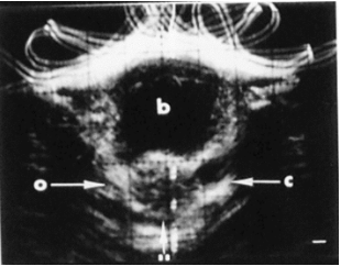
Figure 1: First generation grayscale sonogram showing the right ovary (o), left corpus luteum (c) and bladder (b), circa 1974.
Another major technical improvement occurred early in my radiology training with first-generation real-time imaging. Ultrasound, at the time, was the first truly cross-sectional imaging modality before CT became widely available. Subsequent advances using ultrasound included color Doppler sonography, 3D volumetric ultrasound, and microbubble-enhanced sonography of vascularity in normal and neoplastic vascular networks (Figures 2 and 3).
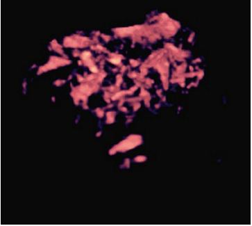
Figure 2: 3D color Doppler sonogram of vascular network within malignant uterine polyp. Note the irregular branching and caliper of the vessels, circa 1992.
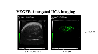
Figure 3: Static images of implanted tumor with green (right) indicating labelled microbubble to VEGF receptor, circa 2002.
Parallel to these diagnostic advances, therapeutic applications of ultrasound developed into many clinical applications. Labelled microbubbles can be directed to tumors for theragnostic purposes (Figure 4). Focused ultrasound can enhance drug delivery, especially when it involves opening the blood brain barrier.
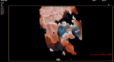
Figure 4: Surface rendition of uterus tube, exophytic fibroid and rectosigmoid colon, circa 2018.
Ultrasound has provided quite a ride for me over the last 50 years or so. I am quite grateful to those who’ve supported, guided and inspired me. Who can imagine what clinical applications will occur during the next 50 years?