In this issue, we talk with Rochelle F. Andreotti, MD, FACR, a Professor of Clinical Radiology and Radiological Sciences and Professor of Clinical Obstetrics and Gynecology at Vanderbilt University Medical Center, about the Ovarian-Adnexal Reporting and Data System (O-RADS™) and its impact on improving women’s health.
Q. Tell us a little bit about O-RADS and its importance to improving quality and safety?
A. O-RADS is one of nine published ACR reporting and data systems for quality and safety. It functions as a quality assurance tool and clinical support system for standardized description of ovarian/adnexal pathology and its management. The system includes a common lexicon that radiologists can use to categorize all levels of risk and assign associated management strategies.
There is a critical need for O-RADS. Too much unnecessary surgery is performed for benign ovarian/adnexal lesions. Most of these lesions are physiologic, non-neoplastic or benign neoplasms; and ovarian cancer is lethal, but rare. Surgery associated morbidity has also been reported to be as high as 15%. On the other hand, initial surgery is frequently performed by a general gynecologist when there are much better outcomes with malignancy when management is by a gynecologic oncologist.
For all of these reasons, we need guidelines to manage benign disease conservatively and to know when to refer patients to a gynecologic oncologist when there is significant suspicion of malignancy. This is where O-RADS comes into play: The goal is to optimize ovarian cancer outcomes while minimizing unnecessary surgery in patients at low risk of malignancy.
Q. What has been the journey of the O-RADS Committee to develop these guidelines?
A. O-RADS is an international initiative that has involved extensive collaboration with competing national and international societies. We began in the summer of 2015 developing our mission and membership. Our membership was primarily derived from several major initiatives that prompted our formation, including:
- The Society of Radiologists in Ultrasound (SRU) Consensus Statement, a North American initiative helpful in determining management of cystic lesions.
- The International Consensus, the first collaboration of European and North American management approaches promoting a more conservative, standardized approach while optimizing the referral pattern to a gynecologist when malignancy is suspected.
- Terms and risk stratification models developed by the International Ovarian Tumor Analysis Group (IOTA).
The committee also consisted of members representing national and international-related societies who could contribute to and eventually help promote our system.
As a result, development of the O-RADS guidelines has been a collaborative, multidisciplinary, international effort that brings together two ultrasound approaches:
- The pattern-based approach commonly used in North America.
- European-based IOTA models with the incorporation of the Assessment of Different Neoplasias in the Adnexa (ADNEX) model, a mathematical risk prediction model.
Q. What has been the work of the O-RADS Committee to date?
A. The O-RADS committee comprises two parallel working groups with experts in radiology, gynecology, gynecologic oncology and pathology: one for ultrasound, which is the primary modality, and another for MRI, which is a secondary, problem-solving tool. Both groups have members representing multiple national and international imaging and non-imaging organizations. In addition to chairing the entire committee, I have chaired and have been primarily involved with the Ultrasound Working Group and Dr. Caroline Reinhold has chaired the MRI Working Group.
Our first phase was the development of a lexicon, a practical uniform vocabulary where we describe the imaging characteristics of ovarian and adnexal lesions. In phase two, the lexicon was applied to risk stratification, which is critical for consistent follow up and management. Only O-RADS Ultrasound includes management strategies, although these management recommendations may include an MRI.
O-RADS Ultrasound Assessment Categories and Management Strategies
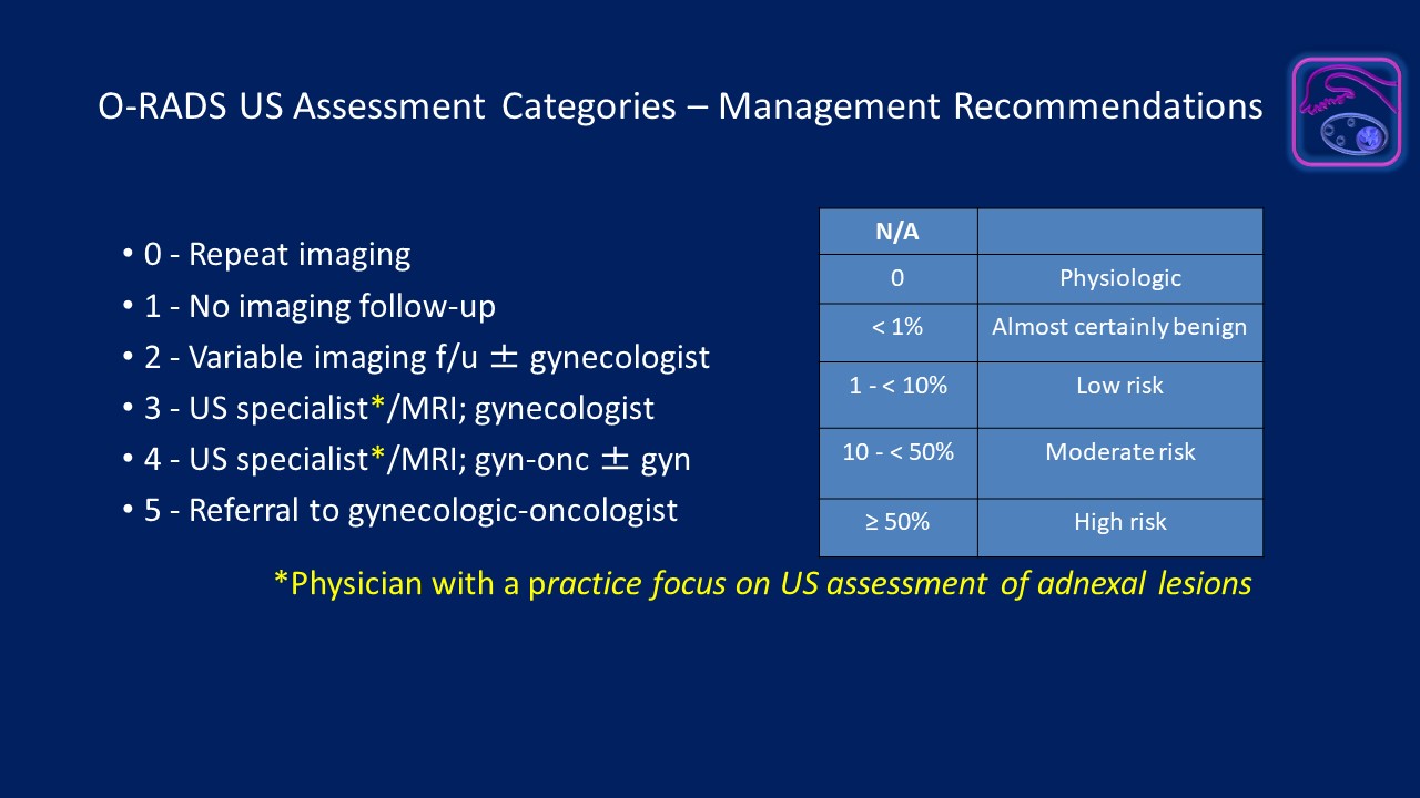
Click the image to view a larger version.
O-RADS Ultrasound Risk Stratification
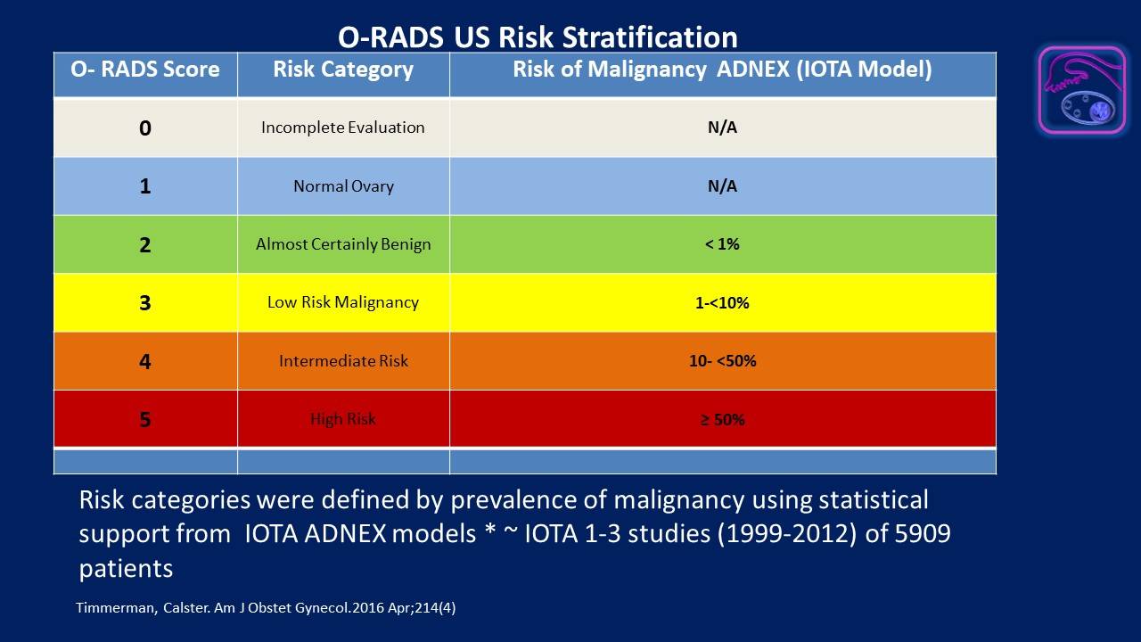
Click the image to view a larger version.
O-RADS MRI Risk Stratification
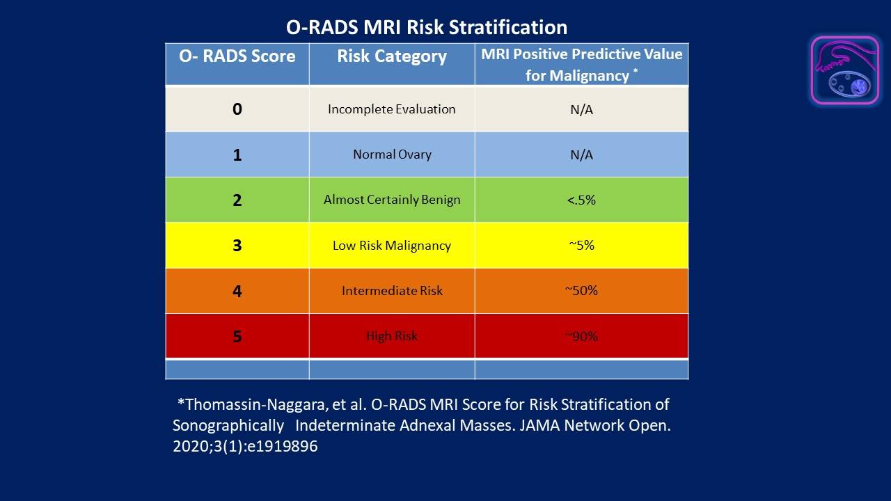
Click the image to view a larger version.
Q. What steps do you recommend that radiologists take to improve quality and safety for ovarian and adnexal mass patient care?
A. First, one must understand that in order to have the highest quality of care, everyone needs to be speaking the same language, which we hope to be the lexicon of O-RADS. This begins with the two general categories of findings that are either physiologic or lesions and continues with the five main subcategories of lesions: unilocular or multilocular with or without one or more solid components and solid or solid-appearing lesions. In addition, a uniform lexicon will permit the collection of reports employing structured tools, providing the opportunity for data scientists to improve outcomes research and ultimately improve ovarian cancer detection rates. Across the board, we all need to use the same terminology.
With terminology in place, we can use the common lexicon to assign the lesion to the correct risk category and recommend appropriate management. Patients with benign-appearing lesions can be offered conservative management, preventing unnecessary surgery. Patients with lesions of higher risk of malignancy in the O-RADS 3 category can be managed by a gynecologist using minimally invasive surgery. Those in O-RADS categories 4 and 5 would need involvement of the gynecologic oncologist for appropriate supervision of care.
Lastly, I am pleased to announce that guidance for the use of O-RADS Ultrasound is now available at the radiologist’s fingertips on the new ACR Guidance App. Simply download the app to your phone or tablet and access the user guide with just a few taps.
Q. What is your hope for the impact that your contributions to O-RADS will make on the specialty and on women’s health?
A. Collectively, the members of the O-RADS Committee feel that this system will have a significant impact on the practice of radiology and women’s healthcare by emphasizing structured classification and reporting of adnexal masses.
As for me personally, any success that I have had in the field of medicine can be attributed to a desire to influence and leave this world, in some way, a little better for it. My hope is that this data system will prove to be something that will make a meaningful contribution and be my legacy to women’s healthcare.